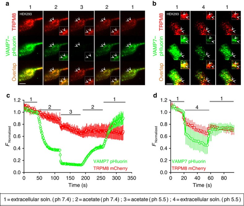
Figure 5. TRPM8 and VAMP7 colocalize in intracellular vesicles and at the plasma membrane.
(a,b) Dual-colour TIRF images showing TRPM8–mCherry and VAMP7–pHluorin co-expressed in HEK293 cells, when perfused with the indicated extracellular solutions. The double arrowheads illustrate the co-expression of TRPM8–mCherry and VAMP7–pHluorin on the cell surface (inset). Scale bar, 10 μm. (c,d) Quantification of total TIRFM fluorescence intensity of TRPM8–mCherry and VAMP7–pHluorin during application of the indicated extracellular solutions. The fluorescent intensities are normalized to the intensity just before switching to solution 2 (c) or solution 4 (d). Data are shown as mean±s.e.m.

