Figure 5.
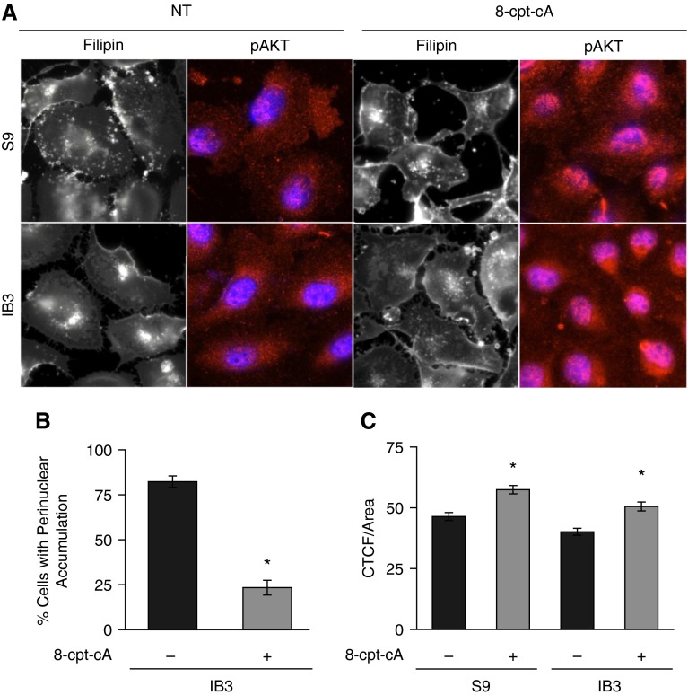
Effect of EPAC1-selective agonist, 8-cpt-cA, on cholesterol distribution in IB3 cells. (A) IB3 cells were either treated with vehicle or with 25 μM 8-cpt-cA for 24 hours and assessed for cholesterol by filipin staining. To ensure efficacy of 8-cpt-cA, cells were immunostained with phospho-AKT (pAKT) antibodies and 4′,6-diamidino-2-phenylindole. Representative images are shown from four separate experiments. (B) Filipin imaging was quantified by assessing cells for perinuclear cholesterol accumulation by a grader blinded to experimental group. Data are presented as percentage of cells with perinuclear accumulation. IB3 cells exhibited 82.3 (±3.2)% of cells with accumulation compared with 23.4 (±6.1)% of IB3 cells treated with 8-cpt-cA (n = 20 separate images for each condition; *P < 0.001). (C) pAKT intensity was determined by assessing cells for corrected total cellular fluorescence (CTCF). Data are presented as intensity (n = 100 separate images for each condition; *P < 0.001). Data represent the mean (±SEM).

