Figure 1.
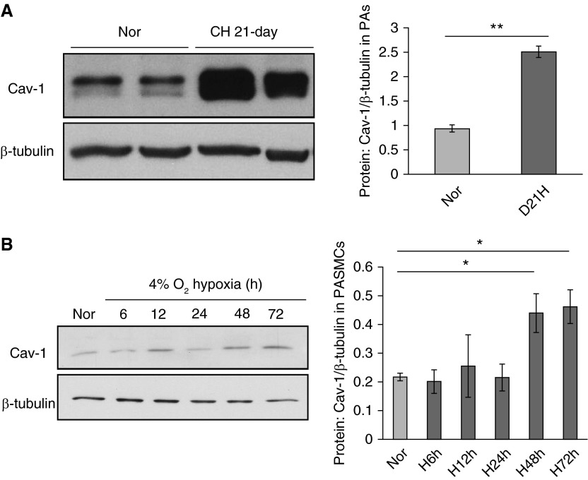
Hypoxic stress up-regulates caveolin-1 (Cav-1) expression in rat distal pulmonary arteries (PAs) and PA smooth muscle cells (PASMCs). (A) Distal PAs isolated from rats exposed to both normoxia (Nor) and chronic hypoxia (CH; 10% O2 for 21 days) were prepared for total protein extraction and Western blot. The left panel shows the blots for Cav-1 and housekeeping protein β-tubulin; the right bar graph represents the quantified data measuring the gray density of Cav-1 and normalized to β-tubulin. (B) Rat distal PASMCs were serum starved and exposed to either Nor or different durations of prolonged hypoxia (Hyp; 4% O2). The left panel shows the blots for Cav-1 and housekeeping protein β-tubulin; the right bar graph represents the quantified data measuring the gray density of Cav-1 and normalized to β-tubulin. Data are presented as means (± SEM); n = 6 in each group. *P < 0.05 versus Nor control; **P < 0.01 versus Nor control.

