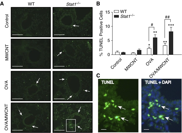
Figure 7.
Stat1−/− mice display significant increases in apoptosis compared with WT mice after OVA sensitization and treatment with MWCNTs. (A) Representative photomicrographs of terminal deoxynucleotidyl transferase dUTP nick end labeling (TUNEL) staining in lung tissue of mice from each genotype and treatment group at 21 days after MWCNT exposure (20× magnification). Arrows indicate TUNEL-positive cells. Scale bars, 500 μm. (B) Quantification of the percentages of TUNEL-positive cells relative to total number of 4′,6-diamidino-2-phenylindole (DAPI)–positive cells 21 days after MWCNT exposure using the Image J protocol described in Materials and Methods. Open bars represent WT mice and solid bars represent Stat1−/− mice. Data are the mean ± SEM (n = 4 animals, control and OVA groups; n = 6 animals, MWCNT and OVA/MWCNT groups). Significant differences compared with controls were determined by one-way ANOVA (*P < 0.05, **P < 0.01, ***P < 0.001). Significant differences between genotypes were determined by two-way ANOVA (#P < 0.05, ##P < 0.01). (C) Higher magnification of inset image in A for Stat1−/− group after OVA/MWCNT treatment showing TUNEL image and merged TUNEL + DAPI. Arrows indicate TUNEL-positive cells. Scale bars, 50 μm.

