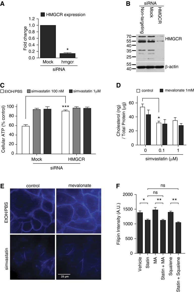
Figure 2.
The plasma membrane cholesterol is reduced by simvastatin. (A) The efficacy of 3-hydroxy 3-methylglutaryl coenzyme A reductase (HMGCR) gene silencing in HBE1 cells was examined by quantitative qRT-PCR. Asterisks indicate significant statistical difference versus matching control with t test. Error bars represent SEM of two experiments. (B) The efficacy of HMGCR gene silencing in HBE1 cells was examined by immunoblotting. The image shown is a representative of three experiments. (C) The gene knockdown of HMGCR in HBE1 cells mimicked the simvastatin-mediated protection against PLY. Asterisks indicate significant statistical difference versus matching control with two-way ANOVA and Bonferroni’s post test. Error bars represent SEM of four to six experiments. (D) The total cholesterol content was analyzed on HBE1 cells upon simvastatin and mevalonate treatment at the indicated concentrations for 24 hours. Asterisks indicate significant statistical difference versus matching control based on one-way ANOVA with Tukey’s post test. Error bars represent SEM of four experiments. (E and F) HBE1 cells were pretreated with 1 μM simvastatin or vehicle control in the presence or absence of 1 mM mevalonate (E and F) or 1 mM squalene (F) for 24 hours. After the medium change, cells were stained with filipin to assess the cholesterol distribution of the membranes. (E) The blue fluorescence staining indicates the filipin probe binding. (F) Quantification data were obtained by fluorimetry using filipin-stained HBE1 cells grown in 96-well microplates. Asterisks indicate significant statistical difference versus matching groups with one-way ANOVA and Tukey’s post test. Error bars represent SEM of three experiments. A.U. arbitrary units; MA, mevalonate; siRNA, small interfering RNA. *P < 0.05; **P < 0.01; ***P < 0.001.

