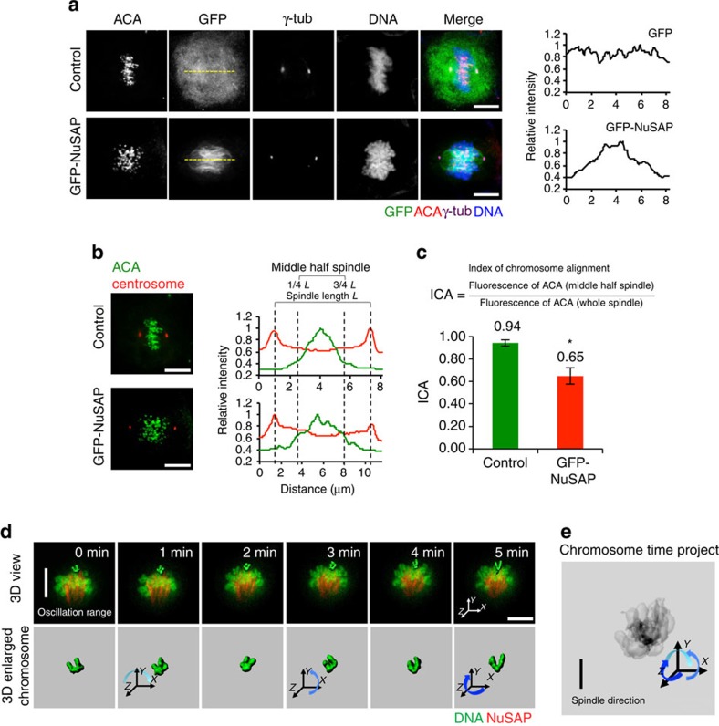
Figure 1. NuSAP regulates chromosome alignment and orientation during metaphase.
(a) Fluorescent images of synchronized metaphase HeLa cells expressing GFP-NuSAP and GFP-vector (control). The line profiles of the GFP channel are presented in the graph on the right. Mitotic spindles were labelled with anti-γ-tubulin and anti-CREST, and DNA with Hoechst 333342. Scale bar, 5 μm. (b) Representative images using the ICA method to analyse centromere alignment in HeLa cells expressing GFP-NuSAP and GFP-vector only (control), with data represented as a line graph on the right. The distribution of ACA fluorescence within the spindle region was measured along the spindle axis following staining with anti-γ-tubulin and anti-Crest. (c) ICAs in HeLa cells expressing GFP-NuSAP and GFP-vector only (control) were analysed as in b. Data were collected from three independent experiments and error bars represent±s.d. *P<0.001, by t-test. (d) 3D time-lapse imaging and 3D single chromosome surface of chromosome oscillation in synchronized metaphase mCherry-H2B HeLa cells stably expressing GFP-NuSAP. Changes in the chromosome orientation are indicated by arrows. Scale bar, 5 μm. (e) Time project image representing the single chromosome misorientation.

