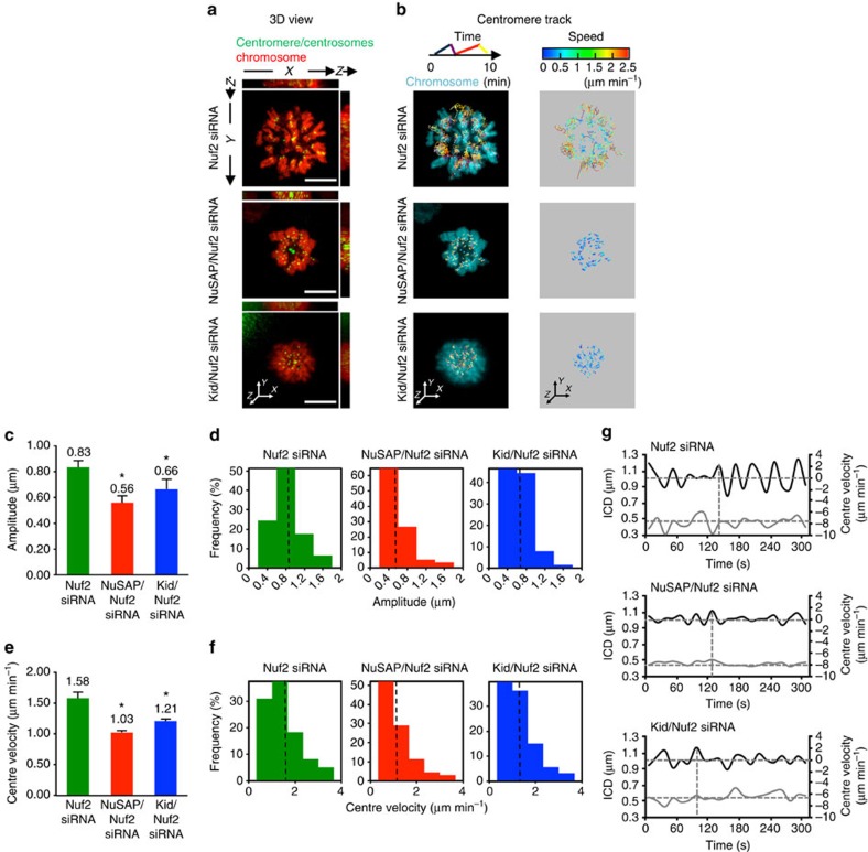
Figure 6. NuSAP tunes the polar ejection force with Kid at interpolar microtubules in monopolar cells.
(a) Representative images of the 3D view using xz and yz projections in Nuf2-, NuSAP/Nuf2- or Kid/Nuf2-depleted monastrol-treated monopolar HeLa cells stably expressing mCherry-H2B. Centromeres were marked with GFP-CENPA and centrosomes with GFP-centrin. Scale bar, 5 μm. (b) Representative images of 3D centromere tracks colour-coded for time and velocity. (c,d) Bar chart and histogram representing the average (c) and the distribution (d) of the amplitude of centromere oscillation in Nuf2-, NuSAP/Nuf2- or Kid/Nuf2-depleted monopolar cells. Error bars represent +s.e.m. *P<0.001, by t-test. (e,f) Bar chart and histogram representing the average (e) and the distribution (f) of the centre velocity. Error bars represent +s.e.m. *P<0.001, by t-test. (g) Representative velocity (black line) and ICD (grey line) versus time plots of the projected centre in a sister centromere pair in the control, NuSAP- or Kid-depleted monopolar cells. The sign of the velocity indicates the direction of centromere movement. The average of the ICD (horizontal) and the minimum (horizontal) and maximum velocities (vertical) are indicated by the dashed lines.

