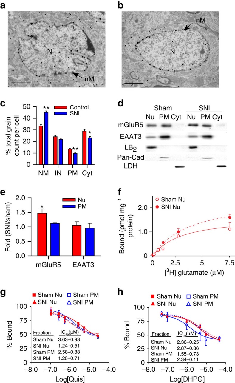
Figure 3. Nerve injury increases SCDH nuclear mGluR5.
Electron-micrographs showing increased nuclear mGluR5 in a SCDH neuron of spared-nerve injury (SNI) (b) versus sham rat (a). (N, nucleus; nM, nuclear membrane). Scale bar, (a,b) 2 μm. (c) Percentage of mGluR5-labelled grains on plasma (PM) or nuclear (NM) membranes, or within cytoplasm (Cyt) or the intranuclear (IN) compartment in SNI and control rats (103 somata were counted from three SNI rats and 86 somata were counted from two control rats, ANOVA *P<0.01; **P<0.001). (d) Western blot of nuclear (Nu), cytoplasmic (Cyt) and plasma membrane (PM) fractions from sham and SNI lumbar SCDH. mGluR5 is increased in nuclear (Lamin-B2 (LB2)) but not PM (Pan-cadherin (Pan-Cad)) or Cyt (lactate dehydrogenase (LDH) fractions from SCDH of SNI rats, quantified in (e). (e) Data shown represent the mean of three experiments, Student's t-test P<0.05. (f) There are significantly more 3H-glutamate sites in SNI nuclear preparations (dashed line) with respect to sham nuclei (solid line) (SNI Bmax=2.87±0.15 pmol mg−1, sham Bmax=1.96±0.21 pmol mg−1, *P<0.05) (g,h) percentage binding and IC50 values of Quis (g) or DHPG (h) on nuclear or PM are comparable in sham and SNI animals. Data shown represent the mean of three experiments, comparisons with Student's t-test. All values in figure are expressed as mean±s.e.m.

