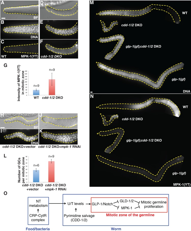
Figure 5.
MAP kinase MPK-1 is activated in the mitotic zone of cdd-1/2 DKO worms fed low-U/T food. (A–F) Representative bright-field (BF) (A,D), DAPI staining (DNA) (B,E), and anti-activated MPK-1 staining [MPK-1(YT)] (C,F) images of dissected gonads from worms of the indicated genotypes. Bar, 5 µm. (G) Bar graph representing the pixel intensity quantification of MPK-1(YT) staining within the whole mitotic zone. (n) Number of gonad arms examined. (H–K) Representative bright-field (BF) (H,J) and DAPI staining (DNA) (I,K) images of dissected gonads from worms of the indicated genotypes, showing that reduction of MPK-1 activity by RNAi partially and significantly increased germline proliferation in the distal end of the gonad arm. (L) Bar graph of statistical data (number of total germ cells within the mitotic zone per gonadal arm) for the animals tested in H–K. (n) Number of gonad arms examined. (M,N) Representative DAPI staining (M) and MPK-1(YT) staining (N) images of dissected gonads from worms of the indicated genotypes, showing that the level of activated MPK-1 is inversely correlated with the mitotic proliferation in the distal end of the gonad arm. An asterisk marks the distal end of the gonad. Bar, 5 μm. (O) Proposed model of the modulation of mitotic germline proliferation by U/T level changes in worms. Both the CRP–CytR complex in food (bacteria) and CDD-1/2 in worms have a great impact on U/T levels; the GLP-1/Notch receptor, a known germline proliferation master regulator, is one critical factor that mediates this protective regulatory system.

