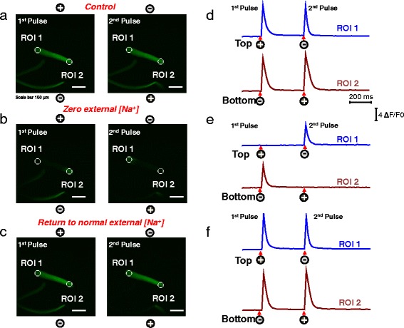
Fig. 4.

Myofibers with uniform responses challenged with a Na+-free extracellular solution exhibit local ALT Ca2+ transients that reverse to UNI responses upon return to normal Na+ containing external solution. Myofibers with uniform responses were loaded with rhod-2 and their Ca2+ transients elicited by electrical stimulation were recorded (a) before, during the treatment with a Na+-free external solution (b), and after washout and return to normal physiological solution (c). After challenging the myofiber with Na-free solution their Ca2+ transients show local and alternate responses when stimulated with alternating polarity bipolar field stimulation. Upon washout of the Na+ free solution and return to physiological conditions, the uniform behavior is completely restored. Rhod-2 signals were imaged and analyzed as in Fig. 1. See video in Additional file 5 (UNI myofiber challenged with Na+-free solution, and the reversibility of effects of Na+-free solution) for the entire time series. The polarity signs indicate the location of the electrodes and their polarity at any given time. Scale bars in a–c are 100 μm. d–f shows the time course of the rhod-2 fluorescence measured at the ends of the myofibers. Circles labeled as regions of interest (ROI) show ROI 1 (myofiber’s upper end, blue trace) and ROI 2 (myofiber’s lower end, dark red trace) the locations used to measure the time course of the rhod-2 fluorescence. Arrows and signs under the traces indicate both the polarity and the time when the pulses where applied. Vertical scale: ΔF/F0 = 4; horizontal scale: time 200 ms
