Fig. 7.

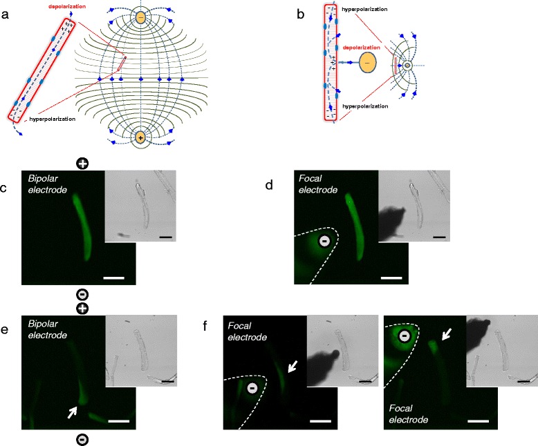
Extracellular electrode configurations used for electrical field stimulation of isolated myofibers and comparison of responses to stimuli by bipolar and local unipolar electrodes. a Theoretical dipole electrical field pattern and isopotential lines generated with two remote Platinum electrodes, separated by about 5 mm and oriented perpendicular to the bottom of the dish. b Theoretical unipolar electrical field pattern and isopotential lines generated with a focal tungsten extracellular stimulating electrode together with a remote field electrode (note: these cartoons represent a simplified approximation to the size, location, and orientation of the electrodes, electrical field, isopotential lines, and current fluxes). c–f Spatial properties of electrically induced Ca2+ transients in UNI and ALT myofibers in response to bipolar or focal unipolar stimulation. In each case, images are confocal snap shots of the time series at the peak of the transient in response to field stimulation (0.5 ms; 15 V/cm). c and d shows spatial properties of electrically induced Ca2+ transient of a UNI myofiber stimulated with a bipolar electrode (c) or with the focal electrode and using the same myofiber (d) (note: the spatial properties of the Ca2+ transients elicited by the bipolar or the unipolar electrode are similar in the UNI myofiber; the signals spread across the entire myofiber regardless of the electrode configuration used). e, f illustrates spatial properties of electrically induced Ca2+ transients of an ALT myofiber stimulated with bipolar electrodes (e) or with the focal electrode (f) either positioned near the center (f left panel) or near the upper end (f right panel) using the same myofiber. Note the differences in the spatial properties of the Ca2+ transients in the ALT myofiber when elicited with remote bipolar or local unipolar electrode. In the case of bipolar stimulation the Ca2+ transient is local and restricted to one end of the myofiber (e); however, when activated by unipolar stimulation, the Ca2+ signal occurs in the myofiber region in close proximity with the location electrode (f). Insets in c–f are transmitted light images to illustrate myofibers and electrode location
