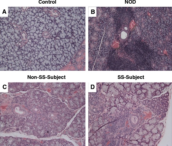
Fig. 1.

Upper panels (a, b) show representative H&E staining of salivary tissues from control and NOD mice while lower panels show representative H&E images for lower-lip biopsies of non-SS subject and SS subject (c, d). As expected, salivary tissue of NOD mouse and lower-lip biopsy of SS subject shows marked leukocyte infiltrations. ×200
