
Fig. 4.
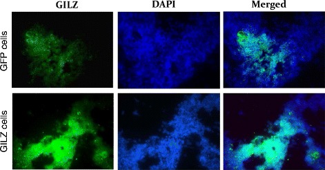
Panels show representative immunofluorescent images for GILZ in control cells (expressing the green fluorescent protein) or in GILZ-expressing cells; DAPI was used as a nuclear marker. ×1000
Official websites use .gov
A
.gov website belongs to an official
government organization in the United States.
Secure .gov websites use HTTPS
A lock (
) or https:// means you've safely
connected to the .gov website. Share sensitive
information only on official, secure websites.
Panels show representative immunofluorescent images for GILZ in control cells (expressing the green fluorescent protein) or in GILZ-expressing cells; DAPI was used as a nuclear marker. ×1000