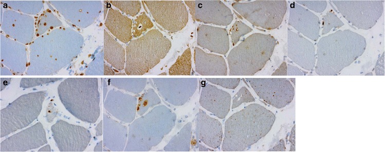
Fig. 4.

a, b, c, d, e. The same patient and muscle as in Fig. 3. The rimmed vacuoles showed material with reactivity for several markers of defect degradation and autophagic processing such as ubiquitin (a), VCP (b), TDP-43 (c), p62 (d) and SMI-31 (e). f, g. The rimmed vacuolar regions of local degeneration filled with autophagosomes, shown by LC3 (f) reactivity, components of mature lysosomes expressing LAMP2 (g)
