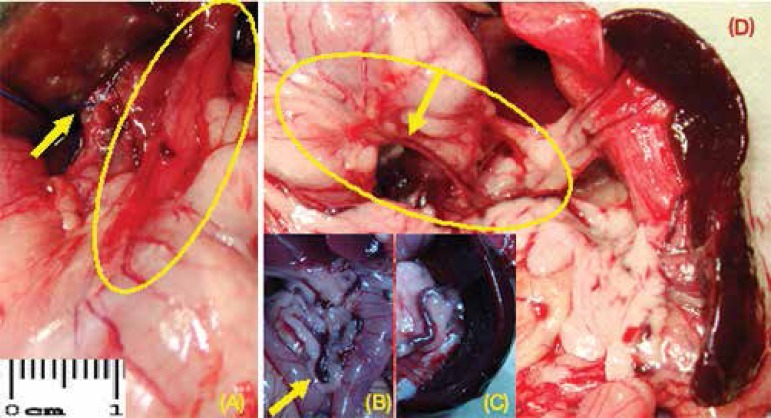
Figure 4.
A) Portal hypertension induced by the partial ligature of the portal vein (arrow) and vasodilatation of the esophagogastric junction - highlighted; B) venous congestion in the territory of the superior mesenteric vein - arrow; C) splenic vein dilatation and tortuosity; D) folded posterior wall of the stomach (highlighted) and dilation of the left gastric vein (arrow), congestion of the splenic hilum vessels, and splenomegaly (animals of group 2)

