Fig. 2.

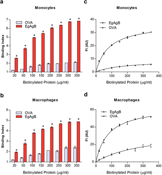
Binding of native EgAgB to THP-1 derived monocytes and macrophages. Binding of native EgAgB to THP-1 monocytes and macrophages was evaluated using biotinylated EgAgB or biotinylated OVA as control (20–350 μg/ml). In (a) and (b) Binding indexes of OVA (grey) or native EgAgB (red) for monocytes and macrophages, respectively; data are expressed as mean values ± SEM of triplicates. Asterisks (*) denote significant differences in binding indexes of native EgAgB with respect to the control (two-way ANOVA followed by Bonferroni’s post-test, p < 0.05). (c), (d) Titration curves obtained for OVA (▲) or EgAgB (●) binding to monocytes or macrophages, respectively. Several fitting models of Graph Prism software were tested and the “binding saturation with one side-specific binding” model showed the best R2 (0.992). The solid line corresponds to the theoretical binding curve obtained for each protein. One representative experiment of three is shown in each panel
