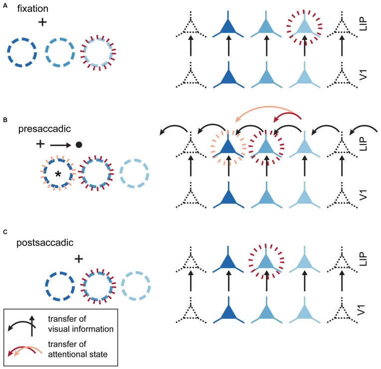
Figure 4.
Visual and attentional remapping. (A) Fixation epoch (prior to saccade plan instantiation). Circles in the left column indicate the position of neuronal RFs; neurons in the right column are colored to indicate corresponding RFs in the left column. Red dashed circles indicate the location of the attentional locus and associated facilitated neurons. (B) Presaccadic epoch (saccade plan instantiated, but not initiated). Black arrows indicate “horizontal” transfer for visual information between neurons triggered by the saccade plan. These transfers are not necessarily instantiated by horizontal connections in cortex, but could equally well reflect changes in the pattern of feedfoward input. The effect of this horizontal transfer is to shift spatial RFs to the right in response to planning a saccade to the right. Two different attentional updating mechanisms are illustrated. (1) Red arrow represents transfer of attentional facilitation as part of the visual remapping signal, which would result in attentional facilitation at the originally attended location (red dashed circle). This is inconsistent with predictive effects demonstrated by Rolfs et al. (2011). (2) Orange arrow: Attentional and visual signals are remapped separately. To generate the observed predictive attentional shifts, attentional state must be remapped twice as far as the visual signal. Importantly, this neuron will not represent the attended location after the saccade is executed, so such a signal would not be useful for stabilizing spatiotopic attention across the saccade. (C) Postsaccadic epoch. RFs revert to their original retinotopic location with attention deployed to the correct spatiotopic location.

