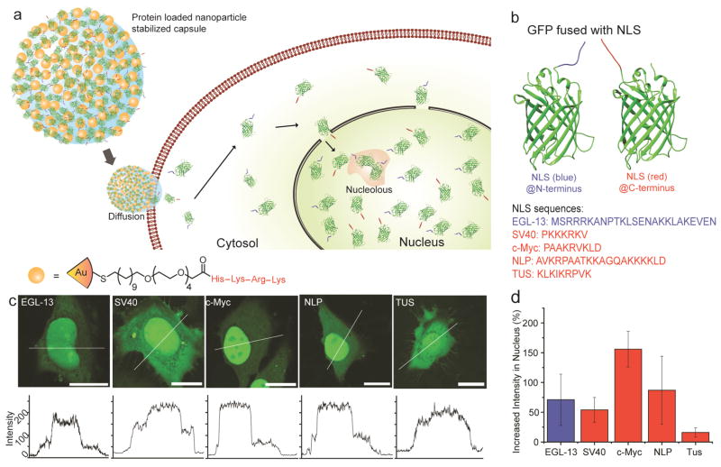
Figure 1.
Delivery of eGFP fused with nuclear localization signals (NLS) to cells using NPSCs. (a) Schematic representing the cytosolic delivery and nuclear accumulation of proteins with NLSs. (b) Structure of eGFP fused with NLSs. (c) LSCM images showing different cellular distribution patterns of eGFP fused with NLSs. Bars: 20 μm. (d) Analysis revealing the increase in nuclear intensities of NLS-eGFPs compared to that in the cytosol of the same cell (6 cells per group).

