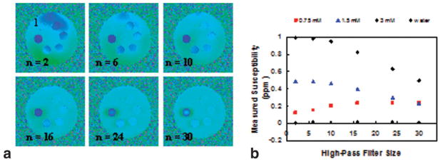
FIG. 3.
a: Series of high-pass filtered phase difference images of the phantom. Field inhomogeneity is reduced with increasing filter size from n = 2 to n = 30. However, contrast is gradually lost among the vials containing different concentrations of Gd with increasing filter size resulting different susceptibility dependence on filter size (b).

