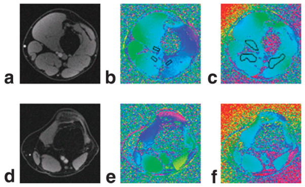
FIG. 5.
a: In vivo magnitude image of an axial slice containing the femoral artery and vein. b: Corresponding phase difference image with shimming. c: Shimming disabled but with weighted least-squares fit correction. d–f: Corresponding axial magnitude and phase images containing popliteal artery and vein.

