
FIG. 8.
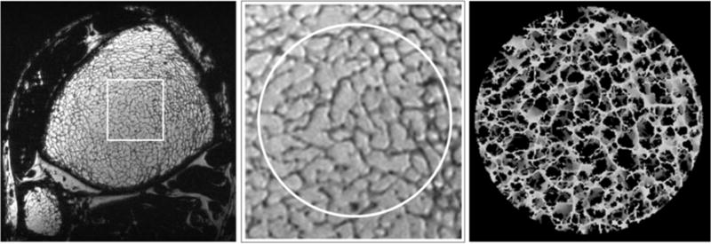
a: Single axial slice in a 3D FSE-OSC image of the distal tibia of a volunteer scanned at 7 T using parallel imaging. Resolution is 137 × 137 × 410 μm3 in a scan time of 10.2 min; (b) magnified view showing high contrast in a region of trabecular bone; (c) 3D rendering of skeletonized core.