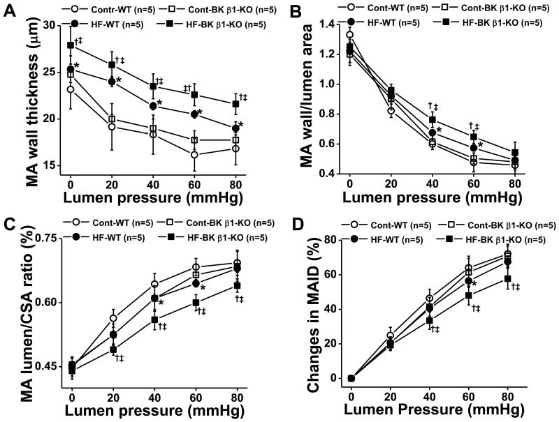
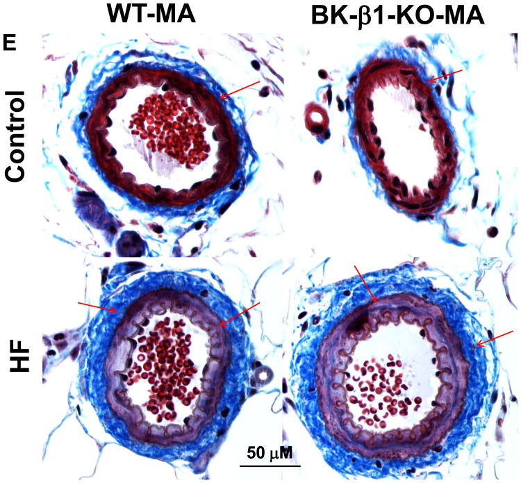
Figure 5.
Comparison of mechanical properties in 24 week diet fed control and HF mesenteric arteries (MA). All measures were taken in Ca2+ free buffer with gradually increased lumen pressure. A), wall thickness, B), wall/lumen ratio; C) lumen/CSA (cross section area) ratio, D) changes in MA ID (MA inner diameter). E, light photomicrographs of control and HF MA. All tissues were stained with Masson's Trichrome stain to highlight perivascular fibrosis (blue-stained connective tissue; arrows). MA in HF fed mice had remarkable vascular remodeling and perivascular fibrosis than vessels from control fed mice, the changes were more predominant in BK β1-KO mice. Images are representative of 6 mice in each group. Data are presented as mean ± SE. *P<0.05, HF-WT vs control WT ; †P<0.05, HF-BK β1-KO vs control BK β1-KO; ‡P<0.05, HF-BK β1-KO vs HF-WT.


