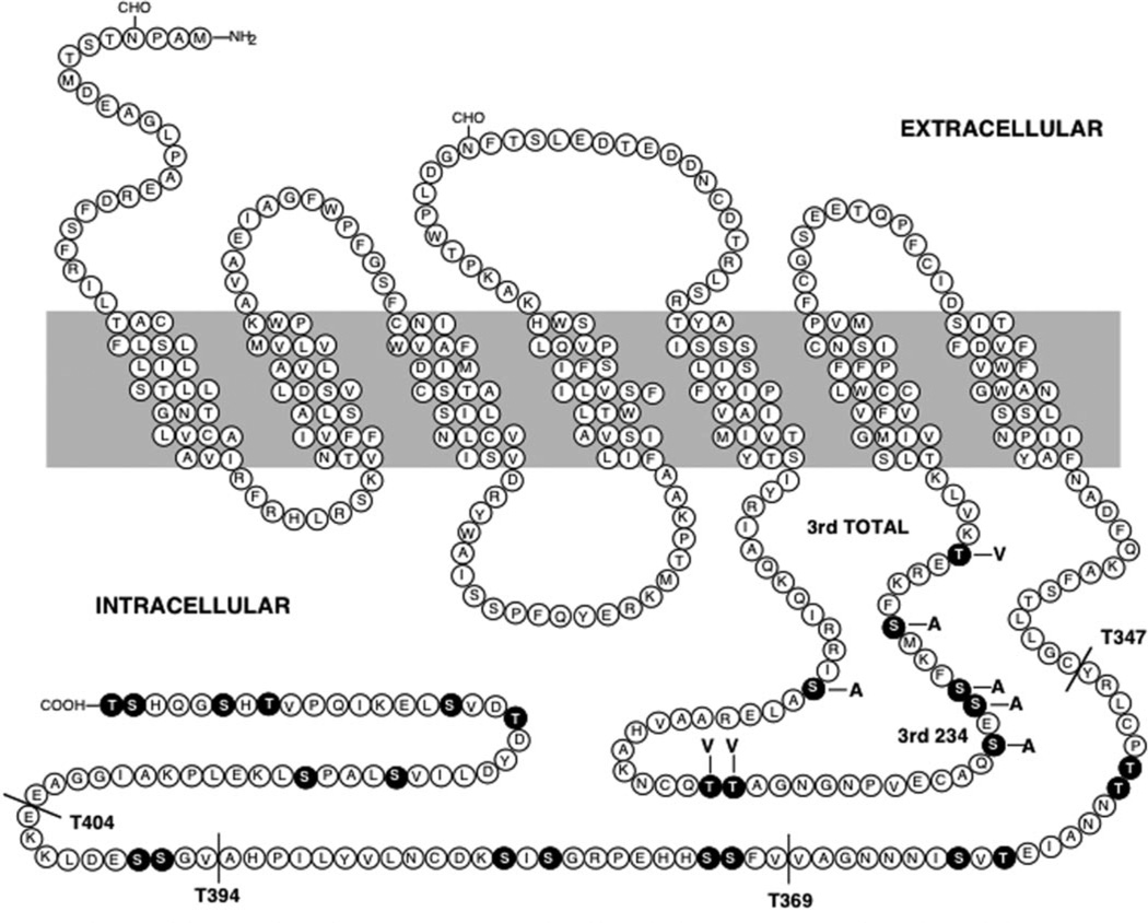
Fig. 1. Diagram of the rat D1 dopamine receptor sequence.
The wild-type (WT) receptor sequence is shown along with the various mutant constructs utilized in this study. The black residues highlight the serine and threonine residues within the 3rd cytoplasmic loop and carboxyl-terminal domains. Four truncation mutants are shown (T347, T369, T394, and T404) in which the receptor was truncated at the position indicated. Two 3rd loop mutants are shown. In the 3rd TOTAL (3rdTOT) mutant, all of the serine and threonine residues within the 3rd cytoplasmic loop were changed to either alanine or valine as indicated. In the 3rd234 mutant, only the three serine residues indicated (serines 256, 258, and 259) were mutated to alanines.

