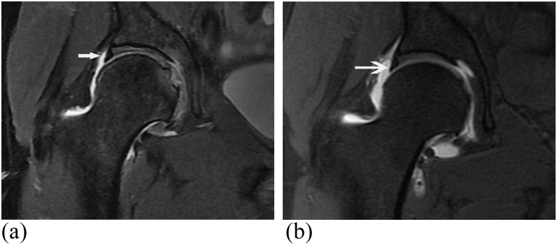
Figure 2.
24-year-old male with hip pain. (a) T2 weighted coronal MR image [3850/55 ms, repetition time (TR)/echo time (TE)] shows acetabular labrum read as normal by both readers (arrow). A tear of the lateral aspect of the acetabular labrum was seen at arthroscopy. No tear was seen on further retrospective review. (b) T1 weighted fat-saturated axial MR arthrogram image (677/12 ms, TR/TE) shows acetabular labrum read as normal by both readers (arrow). A tear of the lateral aspect of the acetabular labrum was seen at arthroscopy. No tear was seen on further retrospective review.

