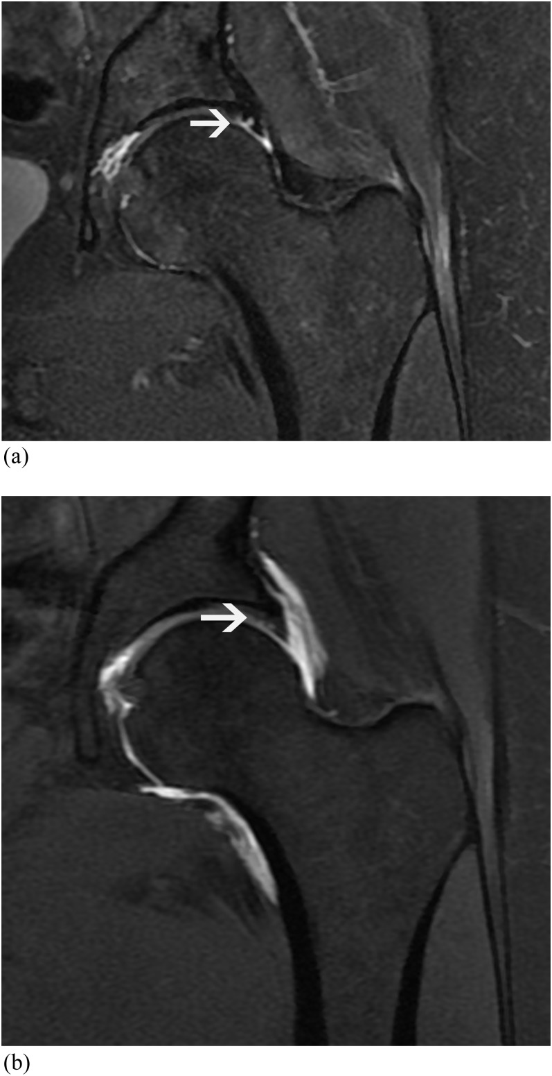
Figure 4.
46-year-old male with hip pain. (a) T2 weighted coronal MR image [3850/55 ms, repetition time (TR)/echo time (TE)] shows acetabular labral tear (arrow). (b) T1 weighted fat-saturated coronal MR arthrogram image (677/12 ms, TR/TE). Acetabular labral tear is less well seen on arthrogram images than on conventional MR images (arrow). Both readers described this as an acetabular labral tear on both MR and MR arthrography. An acetabular labral tear was confirmed on arthroscopy.

