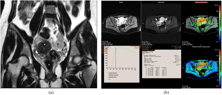
Figure 5.
Right ovarian poorly differentiated Sertoli–Leydig tumour in a 44-year-old female, amenorrhoeic since 7 months. (a) Coronal T2 weighted fast spin echo shows right ovary purely solid pelvic mass of intermediate signal intensity (star). Note that the right ovary shows few follicles. Normal left ovary (black arrow). (b) Quantitative assessment of the right ovarian mass displayed a high maximum relative enhancement percentage of 418%, Tmax of 119 s, and Type III malignant curve pattern seen demonstrated in a collective figure that included T1 high-resolution isotropic volumetric examination source image, subtraction, colour-coded and colour mapping images. C, cervix; R, rectum.

