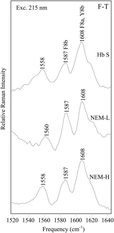
Figure 6.
Comparison of 215 nm-excited UVRR F-T difference spectra. F-T difference spectra of Hb S (top), NEM-H Hb (center) and NEM-L Hb (bottom), were obtained with 215 nm excitation. T-state samples were 1.0 mM Hb, in a 1.0 M potassium phosphate buffer, pH 7.1. Fiber samples were 1.0 mM Hb, in a 0.1 M potassium phosphate buffer, pH 7.1. Fiber formation was initiated by a temperature jump from 10 to 35 °C.

