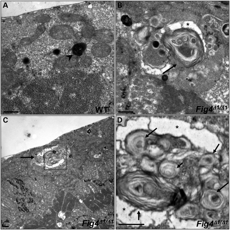
Figure 4.
Ultrastructural analysis of WT (A) and Fig4Δ1/Δ1 mutant (B–D) larval muscles. Boxed area in (C) is magnified in (D). WT lysosomes are small and electron-dense (arrowhead in A), whereas many Fig4Δ1/Δ1 lysosomes are markedly expanded (arrows in B–D) and contain numerous membranous whorls. Asterisk indicates electron-lucent regions that surround membranous whorls. Scale bars: 2 μm.

