Figure 1.
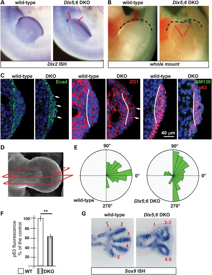
Histology of the AER of limbs of Dlx5;6 DKO mouse embryos. (A) Whole-mount in situ hybridization on WT (left) and Dlx5;6 DKO (right) hindlimbs at age E 11.5, to detect Dlx2 mRNA. Red arrows indicate the beginning and the end of a ‘reduced expression’ region. (B) General appearance of WT (left) and Dlx5;6 DKO (right) hindlimbs, at the age E11.5, in whole-mount bright field examination. Red-dotted lines show the contour of the AER. (C) Immunostaining on sections of WT or Dlx5;6 DKO hindlimbs (respectively on the left and on the right of each pair) to detect E-cadherin (left), ZO-1 (middle), GM130 and p63 (right). While lines indicate the contour of the AER. Arrows indicate cells with clearly expression pattern defect. Scale bars are inserted. (D) Scheme of the section plane. (E) Rosette diagram of the orientation of the Golgi with respect to the surface in WT and Dlx5;6 DKO limbs. (F) Quantification of p63 expression levels in WT and Dlx mutant limbs, expressed in percentage of the WT (=100%). Asterisk indicates statistical significance. (G) In situ hybridization on sections of WT (left) and Dlx5;6 DKO (right) hindlimbs, to detect the Sox9 mRNA. Red numbers indicate the digits, 1 is the thumb.

