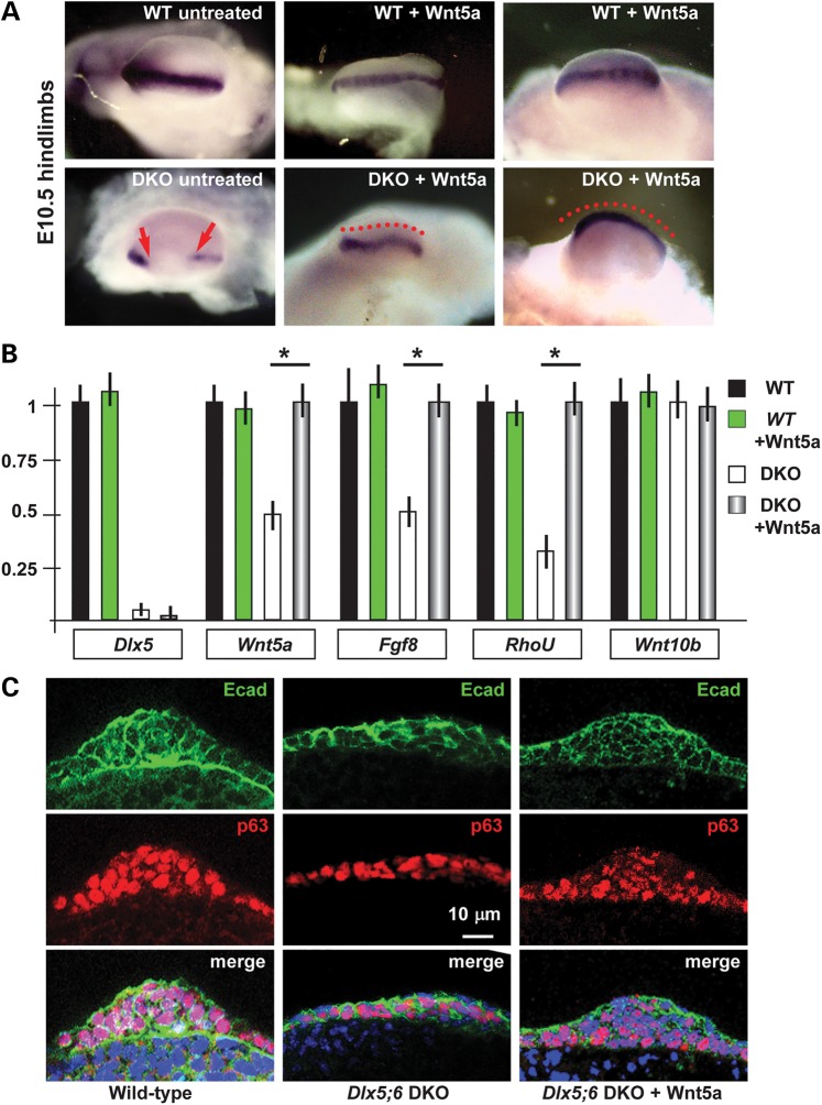
Figure 5.
Cultured normal and Dlx5;6 DKO limbs and their response to Wnt5a. (A) Whole-mount in situ hybridization to detect Dlx2 on cultured limbs. Micrographs are taken from the distal side (AER toward the camera). Red-dotted lines indicate the contour of the AER. Red arrows indicate the beginning and the end of a ‘no expression’ region. (B) Real-time qPCR to determine the relative abundance of Dlx5, Wnt5a, Fgf8, RhoU and Wnt10b, in cultured WT or Dlx5;6 DKO hindlimbs, either treated with Wnt5a or left untreated. Histogram legend is reported on the right. The expression in the untreated WT limbs is normalized =1. Asterisks indicate the statistical significance. (C) Immunofluorescent staining of sections of cultured hindlimbs from WT untreated (left panels) or Dlx5;6 DKO embryos, either left untreated (middle panels) or treated with Wnt5a (right panels). Staining for E-cadherin (green) and p63 (red) are shown. Bottom panels show the merged signal. While lines indicate the contour of the AER. Scale bars are inserted. The section plane is the same as in Figure 1D.

