Fig. S2.
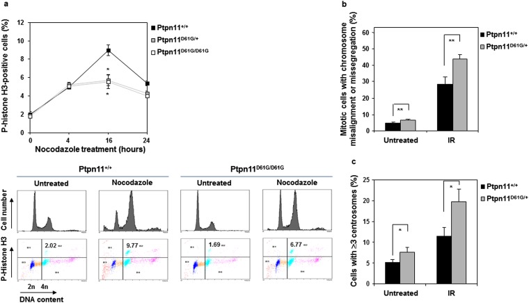
Compromised mitotic checkpoint, aberrant mitosis, and centrosome amplification in Ptpn11D61G mutant primary MEFs. (A) Ptpn11+/+, Ptpn11D61G/+, and Ptpn11D61G/D61G MEFs at passage 3–5 were treated with nocodazole (400 ng/mL) for the indicated times. Cells were collected and the percentage of mitotic cells was determined by PI and p-histone H3 double staining followed by flow cytometric analyses. Representative flow cytometric profiles of cells treated with nocodazole for 16 h are shown in the lower panel. (B) Ptpn11+/+ and Ptpn11D61G/+ MEFs at passage 3–5 were irradiated (10 Gy). Twenty-four hours later, cells were stained with anti–p-histone H3 and anti–β-tubulin antibodies and DAPI. The mitotic cells with aberrant morphology (chromosomal misalignment at metaphase and chromosome missegregation at anaphase/telophase) were counted. More than 100 cells for each cell type were examined. The data shown represent the mean ± SD of the three experiments. (C) Ptpn11+/+ and Ptpn11D61G/+ MEFs at passage 3–5 were irradiated (10 Gy). Twenty-four hours later, cells were immunostained with anti–γ-tubulin antibody, and cells with ≥3 centrosomes were scored. More than 1,000 cells for each cell type were examined. The experiments shown in all panels were performed using three different batches of cell pools. The data shown represent the mean ± SD of three cell pools.

