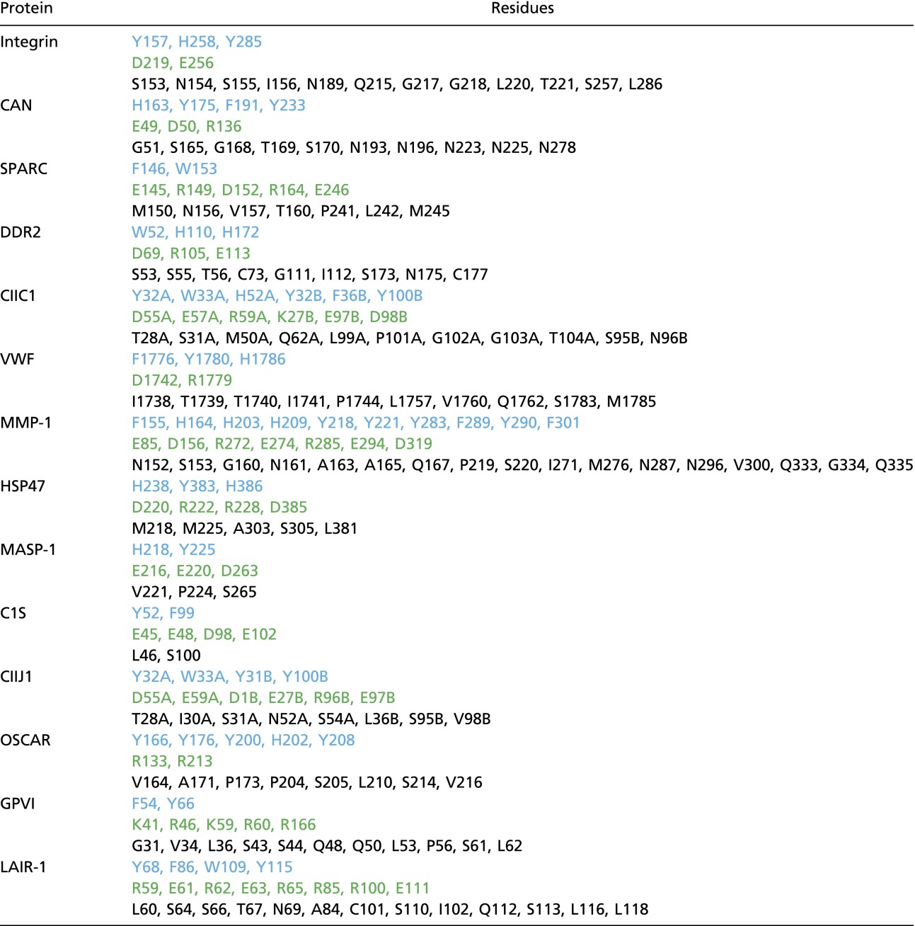
Table S2.
Protein residues that contact CLPs
 |
Protein residues that form contacts under 4.5 Å with a CLP in 11 crystal structures are categorized based on their aromatic (blue), charged (green), or other side chains. In the absence of a crystal structure for GPVI and LAIR-1, the residues found to interact with CLP in NMR or mutational studies are included.
