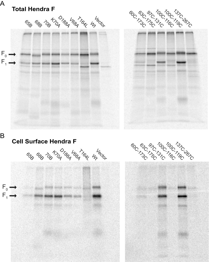
Fig. S3.
Total and cell surface expression of HeV F mutants. (A) Total immunoprecipitated HeV F fraction. Vero cells were transfected with HeV F and HeV G in pCAGGS, and then grown in Cys−Met− medium with Easy-Tag EXPRESS35S Protein Labeling Mix for radiolabeling. Cells were biotinylated with Sulfo-NHS-Biotin and lysed in radioimmunoprecipitation assay buffer. HeV F was immunoprecipitated from the cell lysate with polyclonal anti-HeV F 527–540 peptide serum and protein A agarose. (B) Cell surface HeV F fraction. The biotinylated cell surface fraction was separated with streptavidin agarose from the total immunoprecipitated HeV F fraction.

