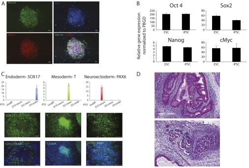
Fig. S4.
The characterization of iPSC5. (A) Immunofluorescent staining of stem cell proteins TRA1-60 and Nanog. Nuclei were stained with DAPI. (B) qPCR of stem genes OCT4, SOX2, NANOG, and c-Myc. (C) In vitro differentiation capability of iPSC5 to three germ-layer lineages. (D) H&E staining of iPSC5 to three germ-layer lineages in teratoma.

