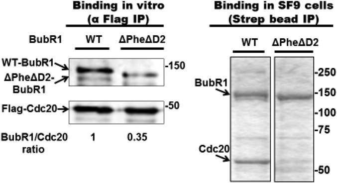
Fig. S2.
Reduced binding of Cdc20 to site 2 of ΔPheΔD2 mutant BubR1. (Left) A total of 500 nM of either WT- or ΔPhe ΔD2-BubR1 was incubated with 300 nM of Flag-Cdc20 for 2 h at 23 °C, followed by immunoprecipitation on anti-Flag beads for 1 h at 4 °C. Subsequently, the beads were washed and subjected to immunoblotting for the indicated proteins. (Right) Insect cells were coinfected with baculoviruses expressing Flag-Cdc20 and either WT- or ΔPhe ΔD2-Strep-BubR1. Complexes of BubR1 were subjected to purification on Strep-Tactin resin as described in SI Materials and Methods, followed by SDS/PAGE and Coomassie staining. Equal amounts of BubR1 were loaded. Numbers on the right indicate the position of the marker proteins (kDa).

