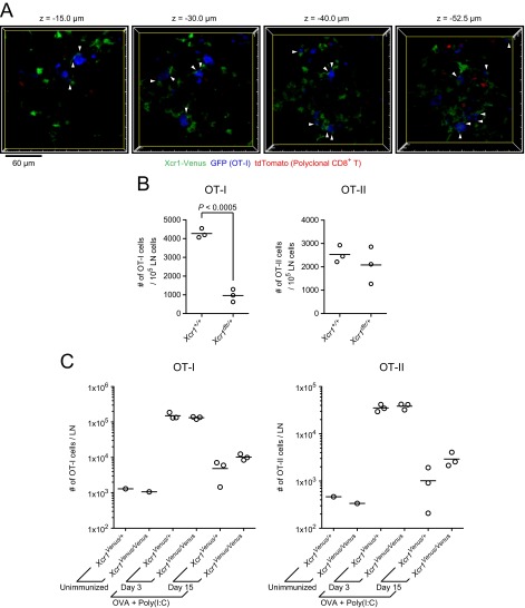
Fig. S2.
Stable interactions of Xcr1-expressing DCs with antigen-specific CD8+ T cells, assessment of the cellular roles for Xcr1-expressing DCs, and assessment of the molecular roles for XCR1 in the CD8+ T-cell response. (A) Single x–y plane fluorescence images of the LN at 20 h postimmunization in Fig. 2A. The arrowheads highlight the stable cell–cell contacts lasting more than 10 min between low-motility OT-I T cells (median velocity ≤4 μm) and Xcr1-expressing DCs (Movie S2). (B) Numbers of transferred OT-I T cells and OT-II T cells in the draining LNs of Xcr1+/+ mice and Xcr1DTRVenus/+ mice. The mice were treated with DT three times, i.e., 1 d before, 1 d after, and 3 d after s.c. immunization with soluble OVA plus poly(I:C). LN cells were analyzed by flow cytometry at 4 d postimmunization. Each symbol represents one mouse. Shown is a representation of similar results from two independent experiments. (C) Totals of 5 × 10⁵ OT-I T cells and 5 × 10⁵ OT-II T cells were cotransferred into Xcr1Venus/+ and Xcr1Venus/Venus mice on day −1. On day 0, the mice were s.c. immunized with 200 µg of OVA plus 20 µg of poly(I:C), and on day 3 and day 15, the draining LNs were analyzed by flow cytometry (unimmunized LNs were analyzed on day 3). Each symbol represents one mouse.

