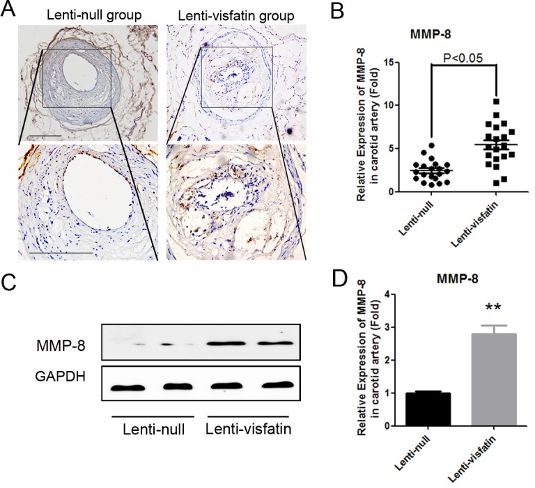
Fig 4. Effects of visfatin on the expression of MMP-8 in vivo.

A, Immunochemical staining of MMP-8 in the plaques in 2 treatment groups is shown. The positive staining areas are shown in brown. B, Quantitative analysis of the results in A (n = 20 in each group). C, Western blot analysis was used to detect the expressions of MMP-8 in the represent samples of carotid plaques from the 2 treatment groups. D, Quantitative analysis of the results in C. Scale bars: 100μm. P<0.05 versus the control group.
