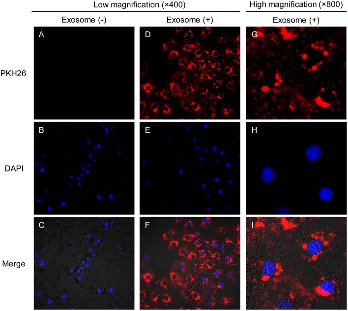
Fig 2. Cellular internalization of OSCC cell-derived exosomes into OSCC cells.
OSC-4 cells were incubated in the presence or absence of 100 μg of PKH26 (red)-labeled exosomes from OSC-4 cells for 4 h and analyzed by confocal microscopy. Nuclei were stained with DAPI (blue). (A-C) Low magnification images of OSC-4 cells without exosomes (400 ×). (D-F) Low magnification images of OSC-4 cells with exosomes (400 ×). (G-I) High magnification images of OSC-4 cells incubated with exosomes (800 ×).

