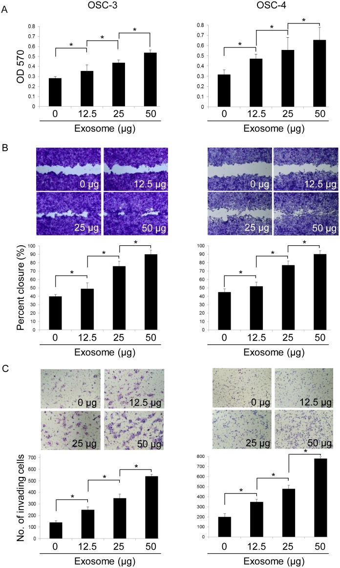
Fig 3. Effects of OSCC cell-derived exosomes on proliferation, migration, and invasion of OSCC cells.
OSCC cell lines were incubated in the presence or absence of exosomes (12.5, 25, and 50 μg) for 24 (A), 10 (B), or 18 h (C). The viable (A), migrating (B), and invading (C) cell numbers were then determined by using the MTT, wound healing, and invasion assay, respectively. The values are presented as the mean ± SD; n = 3 for each group. * p < 0.05 against control OSCC cells, by Mann–Whitney's U-test.

