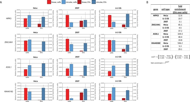
Fig 3. circRNAs are enriched in EV preparations over linear counterparts.
RNA isolated from EV preparations and corresponding source cells was analyzed by real-time quanitative RT-PCR for indicated backspliced circRNAs and the linear spliced mRNA counterparts of the same gene. (A) The relative quantity of each RNA is shown with the level in cells plotted on the primary (left) axis and in EVs on the secondary (right) axis of each bar graph. Error bars are standard deviation of triplicate reactions. (B) Fold enrichment of circRNAs compared to the linear spliced counterpart RNAs in EVs over their corresponding source cells. Fold enrichment is calculated from the qPCR CT (threshold cycle) values for each sample according to the formula shown.

