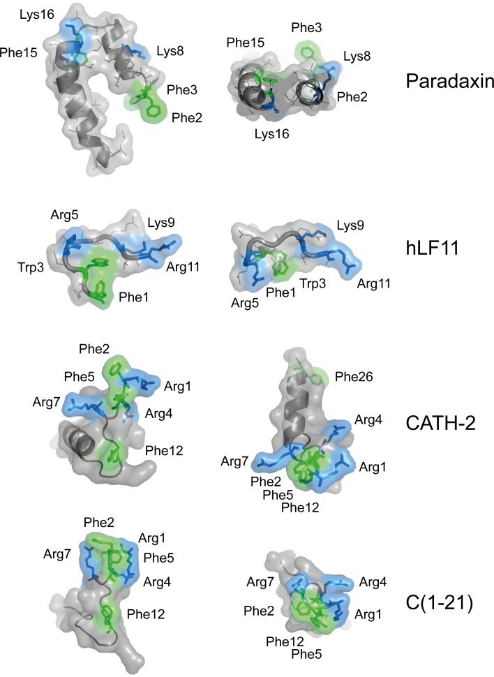
Fig 6. Structural representation of LPS-neutralizing CATH-2 and C1-21 peptide.
Representations of the known 3-dimensional structure of paradaxin and human lactoferrin-derived peptide hLF11, cationic peptides with known LPS-binding affinity and the deduced structures of CATH-2 and C(1–21). Side chains reported or deduced to be in closest proximity to lipid A are depicted in green (aromatic side chains) and blue (basic side chains). Representations were adapted from the published structures (RSCB Protein Data bank; http://www.rcsb.org/pdb/home/home.do). The structure of CATH-2 analog C(1–21) was predicted using iTasser.

