
Fig 4. Morphological changes in heart, lungs and liver tissues.
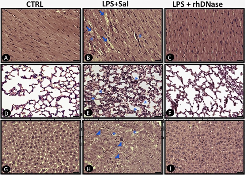
Animals were euthanized 12 h after endotoxic shock induction, and the heart, lungs, and liver were isolated, fixed by immersion in 10% paraformaldehyde, dehydrated and embedded in paraffin wax. Then, 5-μm-thick sections were stained with hematoxylin and eosin for histological examination. The images are representative of heart, lung and liver sections from the CTRL (A, D, G), LPS+Sal (B, E, H) and LPS+rhDNase (C, F, I) groups. Arrows indicate edema. Stars indicate leukocyte infiltration. Arrowheads indicate hyperplasia and hypertrophy of Kupffer cells. n = 5 per each experimental group.