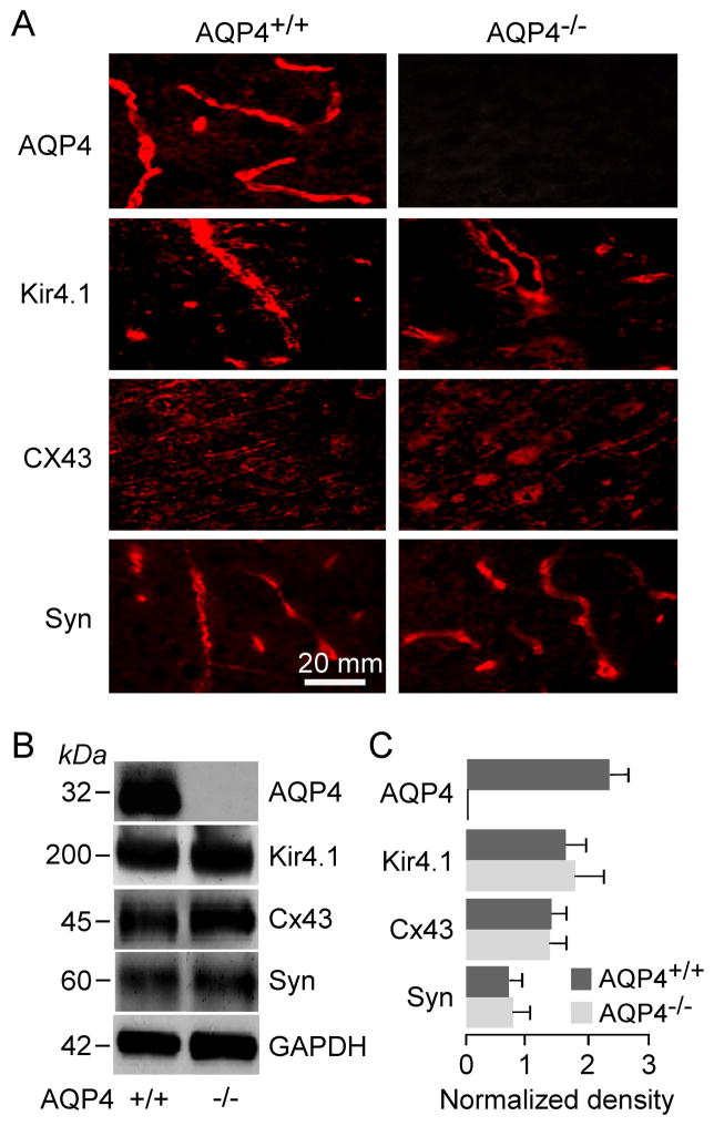
Figure 4.
Expression of AQP4, Kir4.1, Cx43, and α-syntrophin in membrane homogenates of brain cortex of AQP4+/+ and AQP4−/− mice. A. Immunofluorescence using specific antibodies. B. Immunoblot with GAPDH as internal control. C. Kir4.1, α-syntrophin and Cx43 protein quantified by densitometry normalized to GAPDH expression (3 mice per group, mean ± SEM, differences not significant).

