Figure 5.
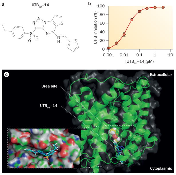
UTBinh-14 inhibition of UT-B. a | The chemical structure of UTBinh-14. b | Concentration–inhibition data for UTBinh-14 inhibition of human UT-B that shows an IC50 of 10 nM when fitted to a single site inhibition model. c | Docking of UTBinh-14 shown in a homology structural model of human UT-B, showing UTBinh-14 binding at the cytoplasmic surface. The computed binding site of urea analogue dimethylurea (yellow) is shown. The inset shows a magnified view of UTBinh-14 bound in a groove at the UT-B channel region. Abbreviations: inh, inhibitor; UT, urea transporter. Permission for part b obtained from the American Society of Nephrology © Yao, C. et al. J. Am. Soc. Nephrol. 23, 1210–1220 (2012).

