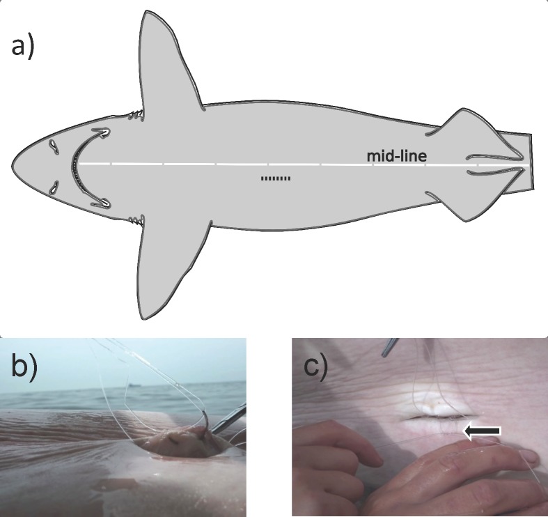
Fig 2. VEMCO Mobile Transceiver (VMT) insertion procedure.
a) Location of the ~5cm incision made just off the midline on the ventral side of the animal. The incision went through the body wall and into the peritoneal cavity. Adapted from [28]. b) Martin Uterine ½ circle reverse cutting needle (size 6) inserted through the serosal surface of the peritoneal cavity, but not protruding through the skin. The non-absorbable nylon monofilament was twice looped through this tissue. c) Example of the surgical knots that were tied to secure the 4 loose ends of nylon monofilament looped through the serosal surface of the peritoneal cavity. Four surgical knots were tied with the two pairs of loose ends and ends were trimmed.

