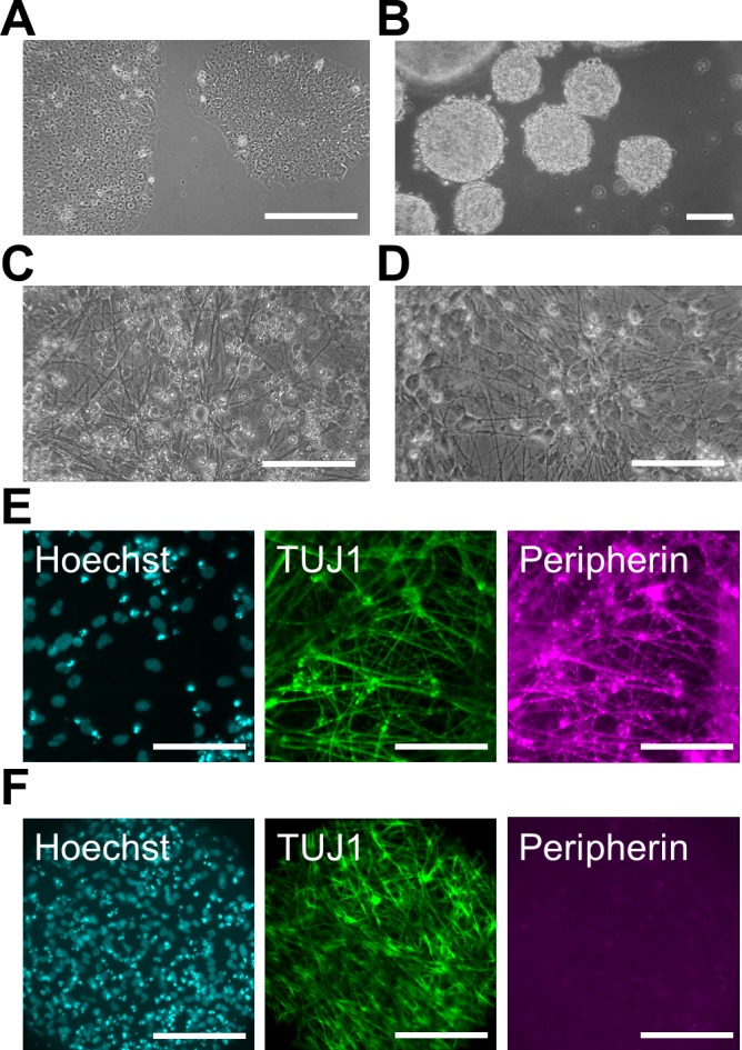
Fig 2. Differentiation of human PNS and CNS neurons.

(A) Phase-contrast image of pre-differentiated human induced pluripotent stem (iPS) cells under a feeder-free condition. (B) Phase-contrast image of embryonic bodies (EBs) before chemical induction on day 0. (C, D) Phase-contrast image of differentiated peripheral nervous system (PNS) neurons (C) and central nervous system (CNS) neurons (D). Induced neurites were detected on day 23 (C) and day 15 (D) in PNS and CNS neurons, respectively. (E, F) Immunofluorescent labeling with antibodies specific for class III beta-tubulin (TUJ1) and Peripherin in PNS (E) and CNS neurons (F) on day 22. Cell nuclei were counterstained with Hoechst 33342. Scale bar: 100 μm.
