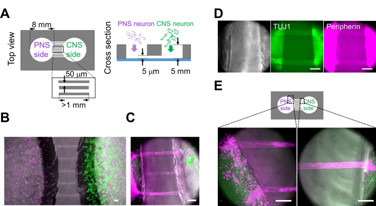
Fig 4. Co-culturing of PNS and CNS neurons in the PDMS chamber device.
(A) Schematic diagram of the polydimethylsiloxane (PDMS) co-culture chamber. Diameter of the chambers was 8 mm. Width of the microtunnels was 50 μm. Although the length of each microtunnel varied depending on its location, the length of each microtunnel was at least 1 mm. The neurites were able to pass through the 5-μm-high microtunnels. (B) Co-cultured neurons close to the microtunnels one day after cell plating. Peripheral nervous system (PNS) neurons were labeled with PKH26 (magenta), and central nervous system (CNS) neurons were labeled with PKH67 (green). (C) Most neurites of PNS neurons passed through the microtunnels 12 days after cell plating. Bundles (magenta) originating from PNS neurons reached the aggregated CNS neurons (green). (D) Phase-contrast images and immunofluorescent staining for class III beta-tubulin (TUJ1) and Peripherin. Neurites that passed through the microchannel originated from PNS neurons, which was confirmed by the expression of TUJ1 and Peripherin (arrowheads) 48 days after cell plating. (E) PNS neurites around the microtunnels 30 days after cell plating. Two different regions, indicated in the diagram, are shown at a high magnification. PNS neurites gathered together and made bundles before and during entering the tunnels. Scale bar: 100 μm.

