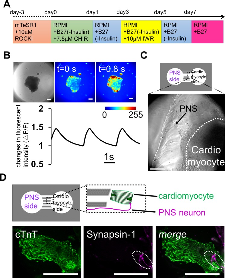
Fig 6. Reconstruction of neuronal networks innervating the heart using iPS cells co-cultured in a microfabricated device.
(A) Schematic diagram of the differentiation protocol for iPS cell-derived cardiomyocytes. (B) Calcium imaging of cardiomyocyte-aggregates on day 16. Contracting embryonic bodies (EBs) showed sustained calcium dynamics. The color bar shows fluorescence intensity, and the change in fluorescence signals confirms that these cell-aggregates contained cardiomyocytes. The graph is showing the kinetics of calcium transients during the recording. (C) Co-culture of peripheral nervous system (PNS) neurons and cardiomyocytes on day 44 after plating PNS neurons. PNS-derived bundles extended from left chamber (arrow) and reached a cardiomyocyte-aggregate, which was in the right chamber (within white dash line). (D) Immunostaining for the cardiomyocyte marker cTnT and the synaptic vesicle marker Synapsin-1 on day 24 after plating PNS neurons. Top, the positional relationship of the axon from PNS neurons and cardiomyocyte. Bottom, localization of Synapsin-1 on a cardiomyocyte (dot line region). Scale bar: 100 μm.

