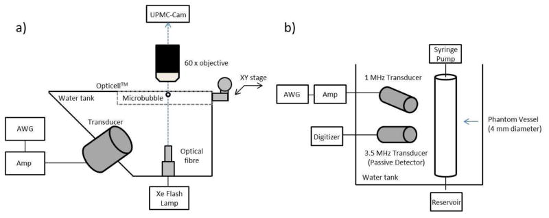
Figure 1.
Schematic diagrams of the experimental apparatuses employed in this study. (a) Ultra-high speed microscopy setup, consisting of a single element 1 MHz transducer, fibre optic light source and Opticell™ phantom within a 2-D translational stage. (b) Passive cavitation detection arrangement, consisting of a 1 MHz transmit transducer and a 3.5 MHz passive cavitation detector transducer. See text for further details.

