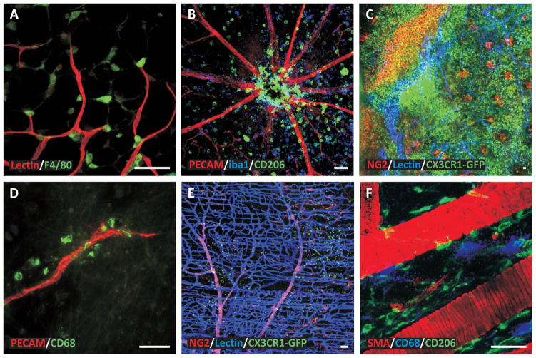
Figure 3. Examples of macrophage spatial distributions during angiogenesis in various tissues.
The heterogeneity in macrophage localization emphasizes the need to identify how and why macrophages are recruited to specific locations within microvascular networks. A) Mouse adipose tissue stimulated by a ligation in a feeding arter (Red: Blood vessels, Green: Macrophages). B) Mouse retina stimulated by IL-1β. (Red: Blood vessels, Blue: M2a Macrophages, Green: Macrophages). C) Mouse ear tissue stimulated by a burn injury (Red: Perivascular cells, Blue: Blood vessels, Green: Macrophages). D) Rat mesentery tissue stimulated by chronic hypoxia (Red: Blood vessels, Green: Macrophages). E) Mouse spinotrapezius muscle (Red: Perivascular cells, Blue: Blood vessels, Green: Macrophages). F) Mouse spinotrapezius muscle (Red: Smooth muscle cells, Blue: Mast cells, Green: Macrophages). Scale bars = 50 microns.

